I found a moss and lichen covered twig on the ground on Boxing Day. Out on a walk with my dogs, the twig caught my attention and I decided to take it home to see what microscopic wonders might be inhabiting it. This blog tells you my main findings.
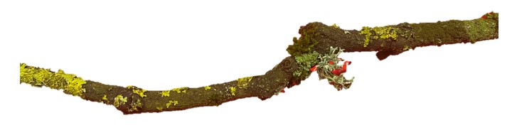
The method itself is an advancement on a standard Moss Safari which is usually a process of sampling and identification. This time I decided to compare the inhabitants of the the three types of habitat on the leaf: yellow crust lichen, grey crust lichen and a tufted moss. Excuse my lack of identification of the habitats at this stage. There was also a forth type of lichen – a grey tufted lichen, but I didn’t have time to sample it.
Introduction
Trees have clusters moss and lichens covering them. Moss Safari often uses moss and lichen to observe the Big Five multicellular organisms (and others) commonly found in these microhabitats. I have often wondered if the microorganisms inhabit particular mosses or lichens preferentially. For example, do tardigrades prefer moss to lichen, or do rotifers prefer to inhabit grey lichen species rather than the yellow lichen species?
In this investigation, I decided to use a rough and ready approach. Take one twig. Isolate the moss and lichens and find out if there is any difference in the number of organisms, the types of organisms and the diversity of organisms.
The null hypothesis is that there is no difference in the number and distribution of multicellular microorganims in the four microhabitats.
Method
Using forceps I pulled the moss from the twig and placed it in a 10 ml vial that contained 2 ml of mineral water (Buxton).
Using forceps to detach the lichen, which involved some pulling and scraping, the two types of lichen were placed in separate 10 ml vials along with 2 ml of mineral water.

The samples were left for 18 hours at room temperature and then sampled.
I shook the lidded vial vigorously and then used a 3 ml plastic Pasteur pipette to draw a 2 ml sample from the vial. I avoided sucking up large lumps of debris.
I then put 4-5 drops of the solution from the Pasteur pipette onto each of five glass ‘dimple’ microscope slides (also known as concave slides). Again, I avoided putting larger pieces of debris onto the slide. Each then had a cover slip placed over the sample in the dimple.

Each slide was observed on a microscope (BMS binocular) with camera (name). With each slide, I took five journeys across the slide in a zig-zag pattern. Each time an organism came into view, it was recorded in a table – just it’s Phylum and a note on it’s appearance: moving, unmoving, dormant, dead etc. Each organism was photographed using the camera. Some active organisms were videoed.
Results
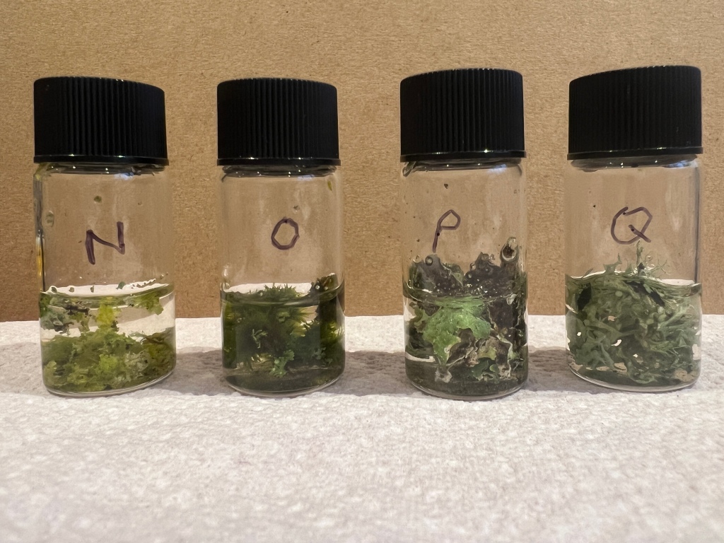
A total of 91 observations (organisms and other items) were recorded from the three microhabitats. Of these 57 were from four of the Moss Safari Big Five (Moss mites (Oribata), tardigrades, rotifers and nematodes), no gatrotrichs were observed. These are displayed in Figure X.
- N – Yellow lichen
- O- Moss tufts
- P- flat grey lichen
- Q- grey forked lichen (results not taken).

The moss had the largest number of organisms living in it, with 34 individuals and four Phyla, then the flat grey lichen had 18 individuals of three Phyla (tardigrades, rotifers and nematodes) and the yellow lichen contained just 7 individuals of the same three Phyla.
Rotifers were the most abundant organism on all three microhabitats, followed by tardigrades, then rotifers. Only one Oribatid (moss mite) exoskeleton was found in the moss sample.
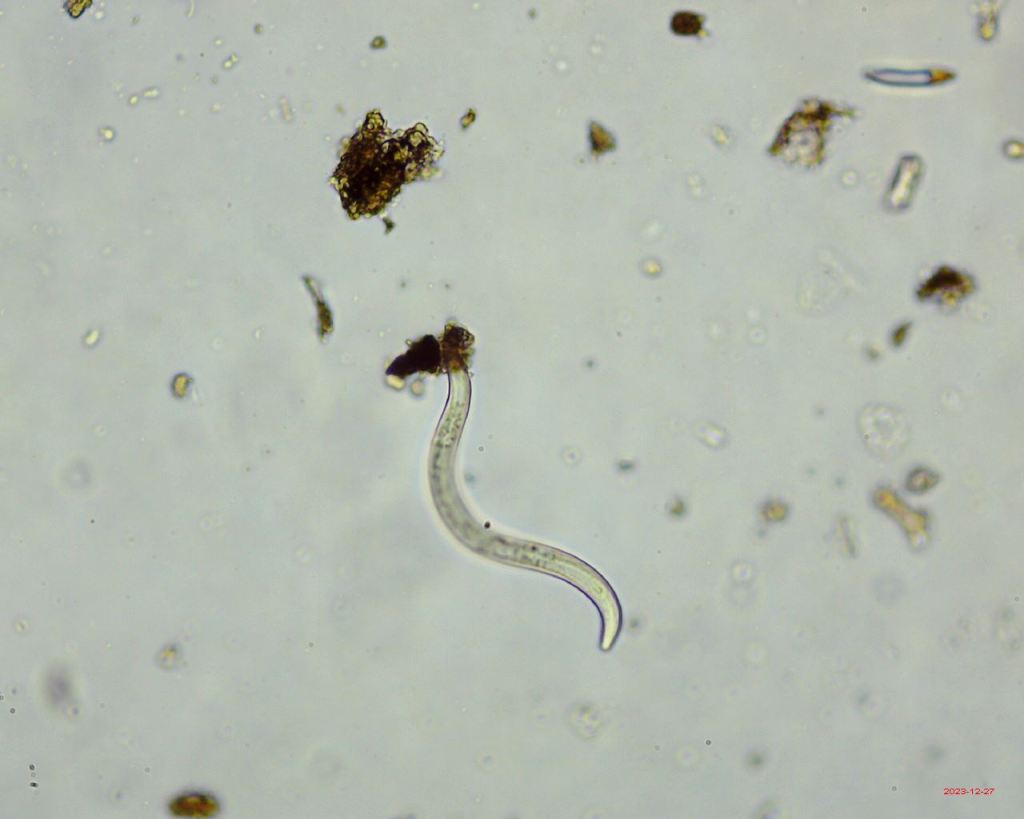
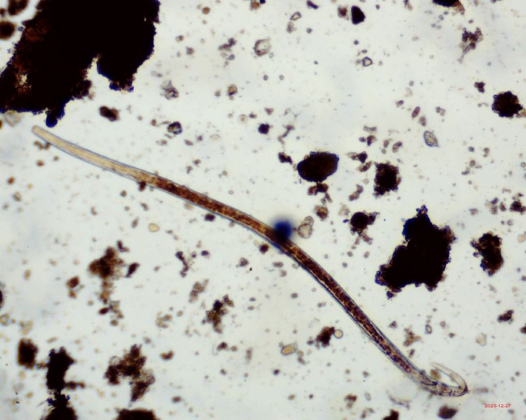
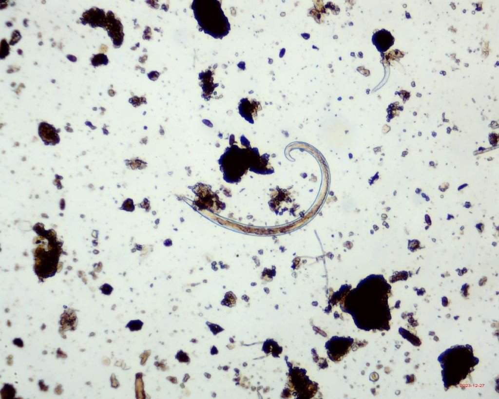
Nematodes
Nematodes at various stages of their life cycle were common in the samples. The images below show a range of juveniles and adults, ranging from just a quarter of a millimetre in length to almost a whole millimetre in length.

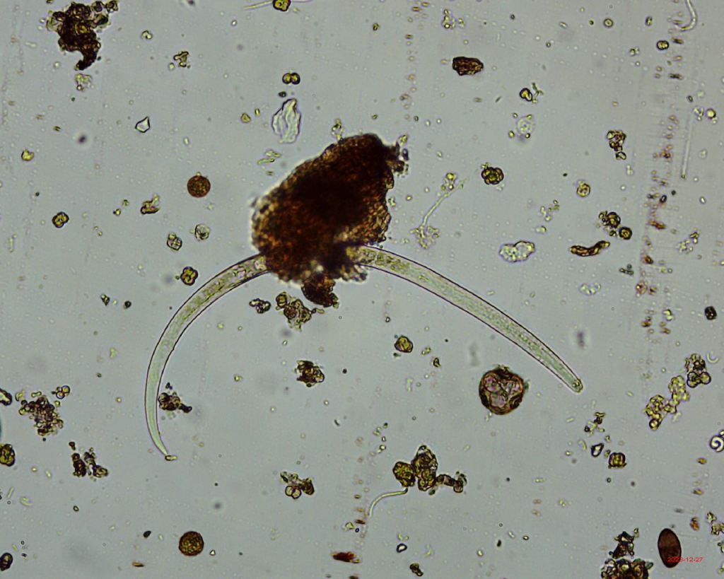


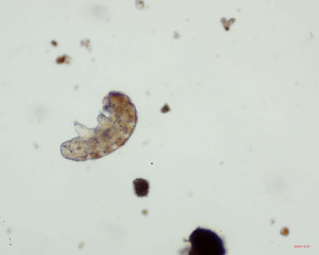
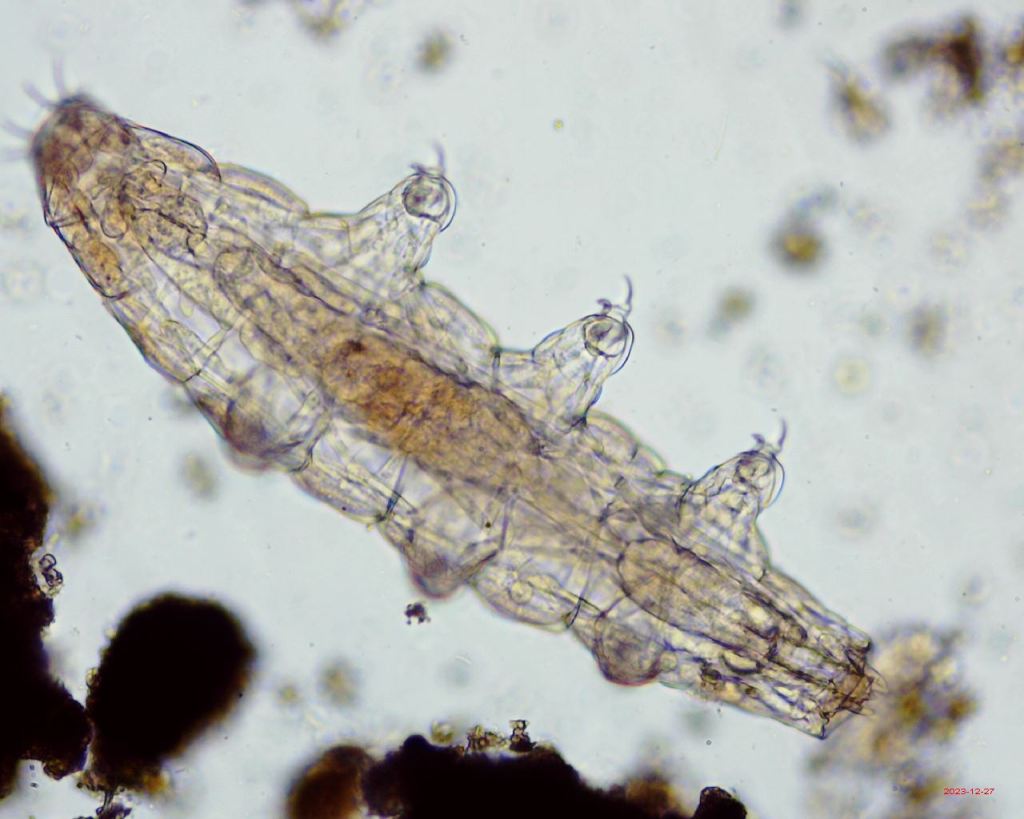
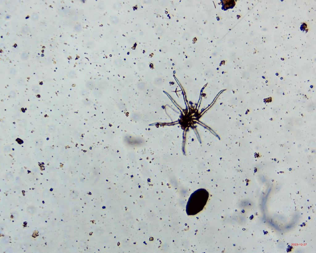
Tardigrades
There were several tardigrades of different genera and species.
These are some examples of them.


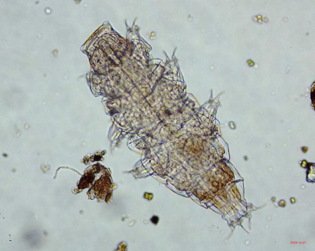

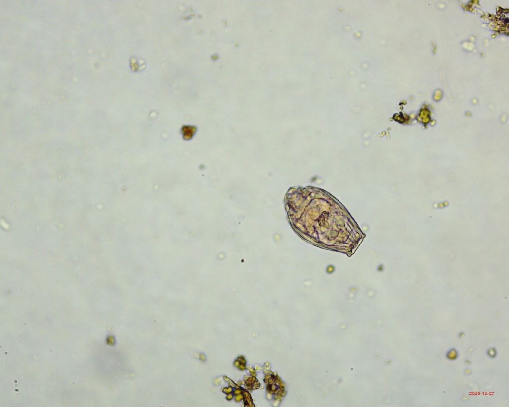
Rotifers
There were a variety of active and contracted Bdelloid rotifers – a selection shown below. These were the most abundant group in all samples.
A. Contracted -possibly dead
B. Eggs inside contracted body?
C. x40 active – walking
D. Possibly going into stasis
E. Contracted but jaws still moving
F. x100 actively walking and feeding
E. Relaxed body, dead decaying. Can see some internal organs.


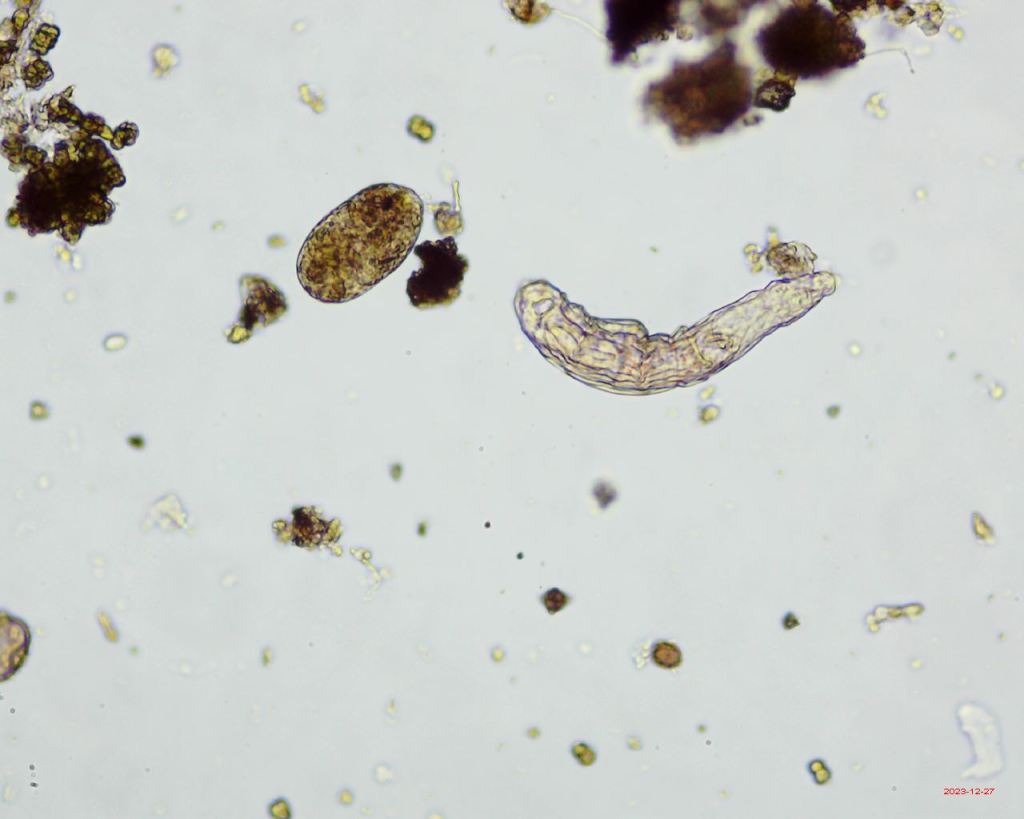

Moss Mite (Oribatid)
I didn’t find any active moss mites, but did find this exoskeleton which suggests that one at least had been present in this moss.

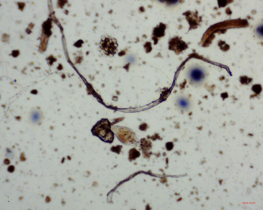
Other findings
The remaining ‘items’ recorded included trichomes (plant gland structures), microfibres, testate amoeba (including Arcella), an unidentified egg and fragments of unidentified arthropod exoskeleton (UMO in the legend below).

Trichomes
Trichomes are glandular appendages found on plants with a variety of functions. I believe that these are what I have found in the samples, in quite an abundance.


I understand that different plants have different trichomes. I am not sure which type of tree the twig came from.
Microfibres
Microfibres and microplastics are commonly found in mosses. Mosses are used to collect microplastics to monitor microplastics pollution. I found three out of the 15 samples I took.
(A) is likely to be a microplastic fibre, (B) is likely to be a microfibre from clothes (may not be a plastic) and (C) is commonly found on microscope slides and maybe from tissues used to clean slides.


I am writing a section of a chapter in the Moss Safari book about microplastics and mosses and their global implications.
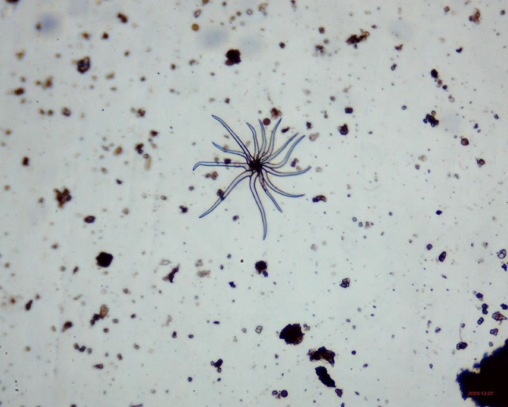
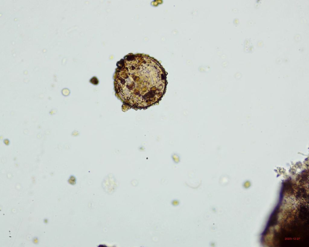
Testate Amoeba including Arcella
I love testate amoeba. These are single cells that grow a shell (a teste) around their bodies for protection (and recent studies suggest some use the testes for active defense). Image B is different from the others as it it is a more spherical species, the others are flat doughnut shapes and are all likely to be Arcella.


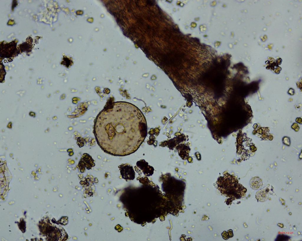
UMO – Unidentified Micro-Organism/Object
I love finding mystery objects. If you know what these are, do let me know.
A. Possibly a piece of arthropod exoskeleton.
B. Possibly a fragment of moss leaf
C. Possibly the jaw and part of the exoskeleton of an arthropod
D. The ‘horn’ shaped, textured object. Absolutely no idea!


Discussion
The null hypothesis was that there is no difference in the number and distribution of multicellular microorganims in the four microhabitats.
The findings suggest that there is a difference in the abundance of multicellular organisms on the twig in the three microhabitats. With moss being home to the most organisms, followed by grey flat lichen and then yellow flat lichen.
I am left wondering why:
Does moss have a greater surface area than the lichen, so more micro-niches?
Does moss hold water for longer, so a preferable habitat?
Is there different chemical composition for each microhabitat? For example, I know yellow lichen indicates high nitrogen content. Does this effect the fauna that live in it?
How much transfer of organisms is there between these habitats on the twig? Do they stay put or actively move? I know that some moss mite species seek out moss to avoid UV radiation and are only temporary visitors. What about the other organisms?
This is a very small sample and would need more samples to see if this was statistically significant. However, as an exercise this is a good example of an experiment that could easily be carried out in schools as part of science club or extra curricular activities.
A small twig with three or four types of habitat does provide a specimen for students to carry out a small investigation. It requires a number of (science curriculum related) skills: identification, ‘fair testing’, sampling, use of a microscope, recording, measuring, presenting data, and analysing data. It is quite easy to do in any school whether inner city or rural. Imagine a whole class analysing one stick, a lot of data could be collected and compared or pairs of students analysing one stick and comparing their results with other student’s sticks.
Are their opportunities for schools from different areas to compare their findings? Could microplastics be compared as well as organisms?
Taking it further
This was a rather time limited experiment done in the space between Christmas and New Year. I don’t always have the time to do these investigations and I certainly did not have the time to do this as detailed as I could.
I could try using a biodiversity index to compare the biodiversity in each microhabitat.
I could attempt some statistical analysis.
I would like to identify the lichens and moss.
I would like to get the genera at least of the tardigrades. I am not sure how easy it is to identify the rotifers and nematodes?
Teachers and enthusiasts
What do you think of this?
Teachers? Could it be used at Key Stage 3, GCSE or Alevel (or equivalent in other jurisdictions)?
Would a resource pack be useful for this?
Enthusiasts. Is there a citizen science project here? What are the opportunities and potential issues?
Let me know your thoughts.
Follow Moss Safari
You can also follow Moss Safari with regular photos, facts and events on social media.
Follow us on Twitter/X, Facebook, and Instagram.
Also now on Threads and Bluesky as @mosssafari.bsky.social (message me if you want a code)

Leave a comment